The idea of removing a brain tumor “through the nose” sounds almost impossible the first time you hear it. But for a specific and growing set of tumors sitting at the base of the skull, this is now one of the most effective and least disruptive ways to operate — no incision on the head, no need to open the skull, and often a faster recovery than traditional surgery. This guide explains what endoscopic skull base surgery actually is, what it treats, what recovery looks like, and how to know whether it might apply to you or a family member.
What is the skull base?
The skull base is exactly what it sounds like — the floor of the skull, the bony platform your brain rests on. It separates the brain above from the structures below and behind your face: the nasal cavity, the sinuses, the eye sockets, and major nerves and blood vessels.
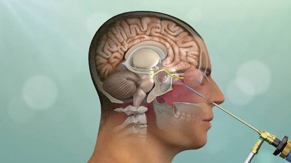
It’s a crowded, delicate neighbourhood. The optic nerves, the carotid arteries, the pituitary gland, and the nerves that control eye movement and facial sensation all pass through or near it. Tumors that grow here are difficult to reach precisely because so many critical structures sit in the way. That’s the problem endoscopic skull base surgery is designed to solve.
What “through the nose” actually means
The medical term is endoscopic endonasal surgery — endonasal simply meaning “through the nose.”
Instead of making an incision in the scalp and removing part of the skull (a craniotomy) to reach a tumor from above, the surgeon uses the nostrils as a natural corridor to approach the tumor from below. There is no external cut and no visible scar.
Here’s how it works in practice:
- The surgeon passes a thin, lighted endoscope — a high-definition camera on a narrow tube — through the nostril into the nasal cavity.
- The nasal passages and sinuses provide a natural pathway toward the base of the skull.
- A small opening is made in the bone of the skull base to reach the tumor.
- Working through the same nostril corridor with specialised long instruments, and watching a magnified live view on a screen, the surgeon removes the tumor.
- The opening in the skull base is carefully sealed — often using the patient’s own tissue — to separate the brain from the nasal cavity again.
Modern skull base surgery is frequently done by a team of two — a neurosurgeon and an ENT (ear, nose and throat) surgeon working together — one navigating the nasal corridor, the other focused on the tumor and the critical structures around it.
What it treats
Endoscopic skull base surgery is particularly suited to tumors that sit in the midline at the base of the skull, within reach of the nasal corridor. The most common include:
- Pituitary tumors (adenomas) — the most frequent reason for this surgery. The pituitary gland sits right above the back of the nasal cavity, making the endonasal route a natural fit.
- Craniopharyngiomas — benign but tricky tumors near the pituitary and optic nerves.
- Meningiomas of the skull base, in selected locations.
- Chordomas and chondrosarcomas — rarer tumors arising from the bone of the skull base.
- Rathke’s cleft cysts and other cystic lesions near the pituitary.
- Certain CSF leaks (leaks of the fluid around the brain) and select sinus tumors extending toward the skull base.
Not every skull base tumor can or should be reached this way — position, size, and how the tumor relates to nearby arteries and nerves all decide whether the endonasal route is the safest option. You can read more about the range of procedures on our skull base surgery page.
Why surgeons choose this approach
When a tumor is well suited to it, the endonasal route offers real advantages over opening the skull:
- No external incision or scar on the head or face.
- No brain retraction — because the surgeon approaches from below, the brain doesn’t have to be lifted or pulled aside to reach the tumor, which lowers the risk of injury to healthy brain tissue.
- A direct, magnified view of midline tumors that would otherwise sit deep and hard to see.
- Often a shorter hospital stay and faster recovery than a traditional craniotomy for a comparable tumor.
- Less post-operative pain and no shaved head or scalp wound to heal.
The key phrase, though, is when a tumor is well suited to it. This is not a “better” surgery in every case — it’s the better surgery for the right tumor in the right location.
What it does not change
It’s worth being honest about the limits, because “through the nose” can sound almost effortless. It isn’t.
Endoscopic skull base surgery is still major surgery on some of the most delicate real estate in the body. It doesn’t remove the need for careful patient selection, it doesn’t guarantee a tumor can be removed completely (some are wrapped around vital structures), and for certain tumors an open approach still gives better, safer access. The nasal route also has its own specific recovery challenges — mainly nasal congestion and the risk of a CSF leak — which we cover below.
The goal is always the safest complete treatment of the tumor, not the smallest possible opening for its own sake.
Recovery: what to expect
Recovery is generally quicker than after open skull surgery, but it has its own rhythm. These are general patterns — your surgeon’s plan takes priority.
- Hospital stay: commonly a few days, depending on the tumor and whether hormone levels need monitoring (for pituitary cases).
- Nasal symptoms: congestion, crusting, and a blocked-up feeling are normal for a few weeks as the nose heals. Saline rinses are usually advised, and you’ll be told to avoid nose-blowing, straining, and heavy lifting early on.
- Headache and fatigue: mild and improving over the first couple of weeks is typical.
- Return to work: many people return to light, desk-based work within a few weeks, though this varies with the tumor and your recovery.
- Follow-up: imaging (MRI) to confirm the extent of removal, and — for pituitary tumors — hormone testing, since the pituitary controls many of the body’s hormones.
The main things to watch for
Two specific issues matter after endonasal surgery, and your team will brief you on both:
- CSF leak — clear fluid dripping from the nose can signal a leak of the fluid around the brain and needs prompt medical attention. Modern sealing techniques have made this much less common, but it’s the complication to know about.
- Hormone changes — after pituitary surgery, hormone levels can shift, sometimes causing excessive thirst and urination (a condition called diabetes insipidus), which is usually temporary and manageable.
Knowing these in advance means you’ll recognise them early and get help quickly if they occur.
Who is a candidate?
Whether the endonasal route is right depends entirely on your specific scan and diagnosis — the tumor’s location, size, type, and its relationship to the arteries and nerves around it. A good skull base team will look at your MRI and CT and tell you plainly whether this approach offers the best combination of safety and complete tumor removal, or whether an open approach would serve you better.
If you’ve been diagnosed with a pituitary tumor or another skull base lesion and are weighing your options, a second opinion from a surgeon experienced in both endonasal and open techniques is worth having — you want someone who can genuinely recommend the right route rather than defaulting to the one they know best.
Frequently asked questions
When performed by an experienced skull base team on a suitable tumor, endoscopic endonasal surgery is a well-established, safe approach. It avoids opening the skull and retracting the brain. As with any surgery, it carries risks — mainly CSF leak and, for pituitary tumors, temporary hormone changes — which your team will discuss with you.
No external scar. The surgery is done entirely through the nostrils, so there’s no cut on the head or face.
Hospital stays are often a few days, and many people return to light work within a few weeks. Nasal congestion and crusting are normal for several weeks while the nose heals.
What tumors can be removed through the nose? Most commonly pituitary tumors, along with craniopharyngiomas, certain meningiomas, chordomas, and some cysts and CSF leaks at the skull base. Suitability depends on the tumor’s exact location and anatomy.
Is this surgery available in Bangalore? Yes. Endoscopic skull base surgery is performed at equipped centres in Bangalore by neurosurgeons trained in the technique, often working alongside an ENT surgeon.
Get an expert opinion in Bangalore
If you or a family member has been diagnosed with a pituitary or skull base tumor, the most valuable first step is a clear assessment of your scan and an honest discussion of the safest approach. Book a Consultation · Send Reports on WhatsApp · +91 63802 71088
This article is for general information and is not a substitute for professional medical advice. Always consult a qualified neurosurgeon about your specific condition.