Medically reviewed by Dr. Shyam D, M.Ch (Neurosurgery), FACS, IFAANS — Consultant Neurosurgeon, Manipal Hospital Electronics City
“You want me to be awake during brain surgery?”
It is the most common reaction when patients first hear about awake craniotomy. It sounds frightening, and it is completely natural to feel anxious. But for the right patient, staying awake for part of the operation is one of the safest ways to remove a brain tumour located near areas that control speech, movement or sensation.
This guide explains what awake brain surgery is, why it is done, what you will feel, how the operation works step by step, who is a good candidate, and what recovery looks like.
What Is Awake Brain Surgery?
Awake brain surgery — medically called an awake craniotomy — is an operation on the brain during which the patient is woken up for a part of the procedure. While awake, the patient answers questions, names pictures, moves their hands or legs, or reads aloud. This allows the surgical team to check brain function in real time while the tumour is being removed.
The patient is not awake for the whole operation. The opening and closing of the skull are usually done while the patient is asleep or deeply sedated. The patient is woken only for the critical part — when the surgeon is working close to important brain areas.
Why Do Patients Need to Stay Awake?
Every person’s brain is wired slightly differently. The areas that control speech, language, movement and sensation are in roughly similar places in everyone, but their exact boundaries vary from person to person — and a tumour can push or shift them further.
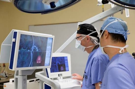
MRI scans, including functional MRI and tractography, give an excellent map of the brain before surgery. But no scan is as accurate as testing the brain directly while it is working.
When the patient is awake, the surgical team can:
- Identify exactly where speech and movement areas are, in that particular patient
- Remove as much tumour as safely possible, right up to the edge of critical areas
- Stop immediately if the patient’s speech or movement starts to change
- Reduce the risk of permanent weakness or speech loss after surgery
In simple terms: the patient’s own responses guide the surgeon’s hands.
Why does removing more tumour matter?
For many brain tumours — especially gliomas — removing as much of the tumour as safely possible is linked to better long-term outcomes. Awake surgery helps achieve the best balance between removing more tumour and protecting brain function.
Does Awake Brain Surgery Hurt?
This is the question every patient asks — and the answer is reassuring.
- The brain itself has no pain receptors. Touching or working on brain tissue does not cause pain
- The scalp is numbed with a local anaesthetic “scalp block”, which works like the injection a dentist gives
- You are asleep or sedated while the skull is opened and closed
- The anaesthesia team stays with you throughout, giving medicines to keep you comfortable and calm
Most patients describe the awake part as strange but not painful. Some feel pressure, hear sounds from the equipment, or feel tired from answering questions. Many patients later say it was far easier than they expected.
How Awake Brain Surgery Works: Step by Step
Before surgery
- Detailed MRI — to locate the tumour and plan the safest route
- Functional MRI and tractography (DTI) in selected cases — to map speech and movement areas and the nerve pathways connecting them
- Pre-operative assessment — speech, language, memory and movement are tested so the team knows your normal baseline
- Practice session — you are shown the pictures, words and tasks you will do during surgery, so nothing is a surprise
- Anaesthesia consultation — to plan sedation and comfort
On the day of surgery
Stage 1 — Asleep or sedated
- You are given sedation or general anaesthesia
- The scalp is numbed with a local anaesthetic block
- Your head is held still in a padded frame
- The surgeon makes the skin incision and opens a window in the skull (craniotomy)
Stage 2 — Awake (brain mapping)
- Sedation is reduced and you are gently woken up
- A speech therapist, neuropsychologist or team member sits near your face and talks to you
- The surgeon uses a tiny electrical stimulator on the surface of the brain. If stimulating a point briefly interrupts your speech or causes movement, that area is marked as critical and protected
- You may be asked to:
- Name objects shown on a screen
- Count numbers or read sentences
- Move your fingers, hand or foot
- Answer simple questions
- The surgeon then removes the tumour while you keep doing these tasks. If any task becomes difficult, the surgeon stops working in that area
Stage 3 — Closing
- Once the tumour is removed, sedation is increased again
- The bone is replaced and the scalp is closed while you rest
The awake part usually lasts around one to two hours, though this varies with the size and location of the tumour. The full operation typically takes several hours.
Technology that adds safety
Awake surgery is often combined with:
- Intra-operative neuromonitoring — continuous electrical monitoring of motor and sensory pathways
- Neuronavigation — a GPS-like system that shows the surgeon’s exact position on your MRI
- Operating microscope — magnified, illuminated view of the brain
- Intra-operative ultrasound — to check how much tumour remains
Who Needs Awake Brain Surgery?
Awake craniotomy is not needed for every brain tumour. It is recommended when a lesion lies in or close to “eloquent” brain areas — regions that control important functions.
Common reasons for awake surgery
- Brain tumours near speech areas — usually in the left side of the brain for most right-handed people
- Tumours near the motor cortex — the area that controls movement of the face, arm and leg
- Low-grade and high-grade gliomas in critical areas
- Some metastases (cancer spread to the brain) close to eloquent cortex
- Epilepsy surgery — to map and remove the area causing seizures while protecting function. Learn more about epilepsy surgery and deep brain stimulation
- Selected vascular lesions such as cavernomas near critical areas
Who is a good candidate?
- Able to understand and follow simple instructions
- Able to communicate clearly before surgery
- Able to lie still for the awake period
- Motivated and well-informed about the process
Who may not be suitable?
- Severe anxiety, claustrophobia or confusion
- Significant speech difficulty already present (testing becomes unreliable)
- Very large tumours causing high pressure inside the skull
- Severe sleep apnoea or breathing problems
- Young children, in many cases — although selected older children can undergo awake surgery with careful preparation
If awake surgery is not suitable, the surgeon may use asleep mapping with neuromonitoring instead. The best approach is chosen individually for each patient.
Benefits of Awake Brain Surgery
- Safer removal of tumours near speech and movement areas
- Lower risk of permanent speech loss or weakness
- More complete tumour removal in many cases
- Real-time feedback that no scan can match
- Often less need for deep general anaesthesia, which some patients tolerate better
- In many patients, a shorter hospital stay and quicker recovery
Risks and What to Expect
All brain surgery carries risks. Your neurosurgeon will discuss how these apply to you:
- Temporary speech difficulty or weakness — common for a few days or weeks as the brain recovers from surgery and swelling settles. Most temporary changes improve
- Seizures during brain stimulation — the team is fully prepared and these are usually controlled quickly with cold saline irrigation and medicines
- Anxiety or discomfort during the awake phase — managed by the anaesthesia team
- Conversion to general anaesthesia — if needed for safety or comfort
- Bleeding, infection or brain swelling — as with any craniotomy
- Permanent deficits — rare, and the whole purpose of awake mapping is to minimise this risk
Recovery After Awake Brain Surgery
| Time after surgery | What to expect |
|---|---|
| Day 0 | Monitoring in the ICU or high-dependency unit. Many patients can talk and move soon after surgery |
| Day 1–2 | Sitting up, eating and walking with support. A post-operative MRI may be done to check tumour removal |
| Day 3–5 | Discharge home for many patients, depending on recovery |
| Weeks 1–2 | Rest at home. Stitches or staples reviewed. Mild headache and tiredness are common |
| Weeks 2–6 | Gradual return to light activities. Speech or physiotherapy if needed |
| After biopsy results | The final tumour report guides whether further treatment such as radiotherapy or chemotherapy is needed |
Tips for a smoother recovery
- Take anti-seizure and other medicines exactly as prescribed
- Do not drive until your doctor clears you
- Rest well, but walk a little every day
- Report new headache, fever, wound discharge, seizures or worsening weakness immediately
- Keep all follow-up appointments and scheduled MRIs
How to Prepare Yourself Mentally
Feeling nervous is normal. These steps help most patients:
- Ask every question — knowing what will happen removes much of the fear
- Do the practice session seriously — familiar tasks feel easier on the day
- Bring a family member to consultations so you both understand the plan
- Tell the team if you feel anxious — sedation and reassurance can be adjusted
- Remember why it is done — staying awake is about protecting the things that matter most: your speech, your movement, your independence
Signs You Should Get a Brain Scan
Awake surgery is only considered after a brain lesion has been found. See a doctor promptly if you notice:
- New seizures, especially in adulthood
- Gradually increasing headaches, often worse in the morning
- Difficulty finding words or understanding speech
- Weakness or numbness on one side of the body
- Changes in personality, memory or behaviour
- Vision problems or persistent vomiting
Read more in 7 Warning Signs of a Brain Tumour You Should Never Ignore and Headache vs Migraine vs Brain Tumour.
Awake Craniotomy in Bangalore — Bangalore Neuro Centre
Dr. Shyam D performs awake craniotomy with intra-operative neuromonitoring for brain tumours in adults and selected children, at Manipal Hospital Electronics City. His practice covers the full spectrum of brain tumour surgery, skull base surgery and complex cerebrovascular surgery.
- M.Ch Neurosurgery — Madras Medical College, with two university gold medals
- FACS — Fellow, American College of Surgeons
- IFAANS — International Fellow, American Association of Neurological Surgeons
- Cerebrovascular fellowship in Japan, skull base training at Weill Cornell, USA, and microneurosurgery training at the Yaşargil course, Istanbul
- Every treatment plan is tailored to the patient’s condition, age, occupation and goals
Patients come from Electronic City, HSR Layout, Sarjapur Road, Bommanahalli, Hosur Road and across South Bengaluru. Learn more about Dr. Shyam or read what to expect at your first visit.
For further reading, see the American Association of Neurological Surgeons and the National Institute of Neurological Disorders and Stroke.
Frequently Asked Questions
No. The brain itself has no pain receptors, and the scalp is numbed with local anaesthetic. You are asleep or sedated while the skull is opened and closed. Most patients describe the awake part as unusual but not painful.
Usually around one to two hours, during the part of the operation when the surgeon is mapping and working near important brain areas. The total operation takes several hours
Get an Expert Opinion on Your Brain MRI
If you or a family member has been diagnosed with a brain tumour, especially near speech or movement areas, an expert opinion can help you understand every option — including whether awake surgery is right for you.
📞 Call / WhatsApp: +91 63802 71088 📍 Manipal Hospital Electronics City, Neeladri Road, Electronics City Phase 3, Bengaluru — 560100
Send your MRI on WhatsApp → | Book a consultation →
This article is for general information only and does not replace a medical consultation. Treatment decisions should be made after a personal assessment by a qualified neurosurgeon.
1 Comment